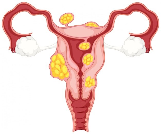
Before we talk about fibroids, it is important that you understand the basics about the reproductive parts in the female pelvis.
Uterus: It is the medical name for the womb and is locates low in the pelvis behind the urinary bladder and in front of the rectum (upper part of back passage). It is a pear-shaped body with smooth muscles in the middle, mucosa (inner wall) and serosa (outer wall).
The endometrium is the inner lining of the uterus which is rich in blood supply and is shed in reproductive life as monthly period.
Cervix is the lower end of the uterus (neck of the womb).
Ovaries: There are two ovaries that release eggs roughly once a month. Ovaries also produce female sex hormones called oestrogen and progesterone.
Fallopian tubes: they are also called egg tunes or oviducts that help to carry eggs from the ovaries to the uterus.
What are uterine fibroids?
Fibroids are tumours that occur within the wall of the uterus. They are also called uterine leiomyomas. They are classifying according to where they are located in the uterus.
- Intramural fibroids: These fibroids are inside the muscular wall of the uterus.
- Sub-mucosal fibroids: these fibroids are inside the lining of the womb. They are called ‘fibroid polyps’ or ‘pedunculated fibroids’ when they get into the uterine cavity.
- Sub-serosal fibroids: They are initially in the muscular wall of the uterus and start growing outside into the pelvis. They are called ‘pedunculated fibroids’ when they are attached to the uterus by a narrow stalk.
- Cervical fibroids: They grow in the cervix.
What causes fibroids?
We don’t know exactly what causes fibroids.
The growth of fibroids is dependent on the amount of oestrogen in the body. So, fibroids develop in the adult women and may increase in size with an advancing age. However, they start to shrink in size after the menopause. They also grow during pregnancy due to the rise of hormones and increased blood supply.
There is a tendency for fibroids to run in families. Women who are obese are also at higher risk of developing fibroids. Furthermore, it has been reported that fibroids are about 3 times more common in women of Afro-Caribbean origin and also that they occur at a younger age.
How common are fibroids?
Fibroids are commonly found in one in three women (30%) of a childbearing age. They are the most common benign (non-cancerous) tumours of the uterus.
Can you have multiple fibroids?
A single fibroid is rare, and it is common to find several fibroids at the time of diagnosis.
They may occur at different sites within the uterus. Fibroids can vary from the size of a pea to the size of a melon.
Are fibroids cancerous?
Generally, fibroids are not cancerous tumours. However, they may become cancerous, but this Is extremely rare.
What are the symptoms of fibroids?
Most fibroids do not cause any symptoms but depending on the number, size and position of the fibroids, you may experience some of the following symptoms:
-Period related symptoms:
- Heavy and/or prolonged periods causing anaemia, social and hygienic problems.
- Irregular bleeding.
- Painful periods.
-Abdominal or pelvic pain.
-Pain during sexual intercourse.
-Abdominal Swelling
-Pressure symptoms: Fibroids grow gradually and large fibroids can start to exert pressure on the bladder and/or the bowel causing:
- Frequent urination
- Bloating
- Constipation(rare)
How are fibroids diagnosed?
Many women do not know that they have fibroids, which may be suspected during a routine gynaecological examination by a doctor. There are some investigations which are helpful to confirm the diagnosis:
-Ultrasound scan of the pelvis: this is a simple procedure involving a lower tummy scan and an internal vaginal scan. A small probe is placed on the tummy to see the picture of the uterus on screen. A small probe may be passe through the vagina for a cleaner image of the uterus as well as the ovaries.
–Hysteroscopy: This procedure may be required sometimes to detect the site and size of fibroids inside the uterine cavity. A thin telescope in passed through the cervix under cavity. A thin telescope to check whether the mass is a fibroid, or a swelling connected to the ovaries or fallopian tubes.
-MRI scan of pelvis: Sometimes an MRI scan is recommended to check whether the mass is a fibroid, or a swelling connected to the ovaries or fallopian tubes.
-Laparoscopy: In this procedure a telescope is inserted inside the abdomen under general anaesthetic to visualise the uterus, ovaries, and fallopian tubes. Occasionally, fibroids are detected during this procedure.
Dr Efterpi Tingi
Consultant Obstetrician and Gynaecologist


